Breaking the Barriers
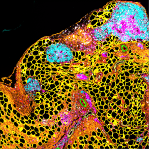
This image offers a glimpse into the tumour microenvironment of metastatic ovarian cancer in a mouse model, caught at a moment of active response to a novel combination therapy that targets both cancer cells and the fibrotic network that supports them. Cancer does not grow in isolation—it reshapes its surroundings, recruiting blood vessels, manipulating immune cells, and remodelling the extracellular matrix (ECM), the fibrous scaffold that holds tissues together.
Here, the therapy is working. Normal fat cells (adipocytes), shown in yellow and typically found lining the gut, are reclaiming space as the tumour recedes. Immune cells—T cells (magenta) and B cells (cyan)—have been recruited from nearby blood vessels (green with a red lining), mobilized to help eliminate remaining cancer cells. Proliferating cells appear in white, marking regions of active cell division.
Threaded throughout the scene is the extracellular matrix, highlighted in orange, composed of structural proteins such as collagen and fibronectin. Once heavily deposited by tumour cells to promote growth and spread, this matrix is now being disrupted as part of the treatment strategy. By targeting not only cancer cells, but also the environment that enables them, this approach reveals a promising way to weaken tumours from multiple angles. Cancer is an ecosystem and restoring balance can help tip the fight in the body’s favor.
Image was acquired by Dr Panoraia Kotantaki, using City of London Centre’s Cell DIVE Imaging Platform, based at the Blizard Institute and in collaboration with Joseph Hartlebury and Dr Florian Laforets.
Visit
What's on
Learn and play
Youth Scheme
Quick links
Website by Hut Six Digital