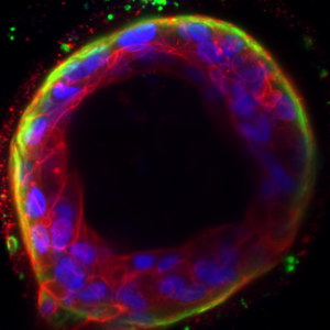
From Cells to Ducts: A Tiny but Incredible Rearrangement in 3D Model
This immunofluorescence image shows how individual primary human luminal and myoepithelial cells (green, CK14) work together in a collagen 3D model to build a tiny two layered tube-like structure (red, phalloidin), similar to the ducts found in a healthy breast. This 3D model helps scientists understand the cell-cell interaction, and the role of the tumour microenvironment in breast cancer initiation and development.
Visit
What's on
Learn and play
Youth Scheme
Quick links
Website by Hut Six Digital