TB: The Master of Deception
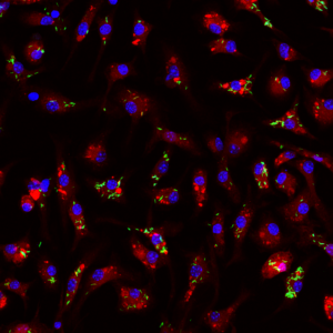
In this image, the tiny green rod-shaped structures are Mycobacterium tuberculosis — the bacteria that cause tuberculosis (TB). They are extremely small, so the picture has been magnified 630 times to make them visible.
These bacteria are living inside immune cells called macrophages, whose normal job is to find and destroy germs. TB is clever: instead of being killed, it hides and survives inside these cells.
The blue structure is the nucleus, the control centre of the cell. The red structures are lysosomes, which are filled with powerful acids and enzymes that usually break down bacteria. Normally, macrophages trap bacteria and deliver them to lysosomes for destruction. In many infections you can see bacteria being digested inside these compartments.
But do you see any TB bacteria inside the lysosomes here (they will look yellow instead of green because of the merging of colours)? No! That’s because Mycobacterium tuberculosis uses special molecules called virulence factors to block this attack. By avoiding the lysosomes, TB can grow safely inside the very cells meant to kill it.
Scientists study this hidden battle to understand how TB survives — and to design better medicines to stop it.
Technical details:
Cells: Bone marrow derived murine macrophages
Bacteria: Mycobacterium tuberculosis H37Rv expressing GFP
Infected macrophages were stained with Lysotracker Red, fixed and mounted using Prolong gold antifade with DAPI to stain the nucleus.
Images were acquired on Zeiss LSM 880 confocal laser scanning microscope using a 63X oil immersion lens.
Visit
What's on
Learn and play
Youth Scheme
Quick links
Website by Hut Six Digital