When Science Finds Love
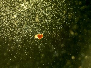
This image, captured using a digital microscope, shows an in vitro culture of CD34 positive cells isolated from umbilical cord blood after two weeks of incubation under hypoxic conditions. These conditions, together with the culture medium, promoted hematopoietic differentiation, allowing the cells to develop into multiple blood cell lineages.
During the culture period, the CD34+ cells generated distinct clonogenic colonies that can be distinguished by their morphology, colour, and spatial organization. In the upper left region of the image, colony forming units–granulocyte/macrophage (CFU-GM) are observed; these progenitors will differentiate into white blood cells involved in immune defence.
In the central area of the image, a burst-forming unit–erythroid (BFU-E) colony is present. These colonies, which give rise to red blood cells, originate from early erythroid progenitors and are characterized by a compact, radially organized structure and an orange-red coloration due to haemoglobin content. In this particular case, against all scientific expectations, the cells appear to have embraced a sense of humour and arranged themselves in a rather lovely shape.
Visit
What's on
Learn and play
Youth Scheme
Quick links
Website by Hut Six Digital