Why does everything else blur when you focus on an object?
By Roberta
In order to understand why this happens we need to take a step back and appreciate the anatomy of the human eye and in particular its primary light sensing component: the retina.
Our eyes are slightly asymmetrical spheres, about an inch in diameter, which consist of:
- A sclera, the white region filled with a clear gel
- The iris, the coloured rings we use to determine our “eye colour”
- The pupils, central black openings that regulate the entrance of light into the eye
- The conjunctiva, a thin layer of tissue at the front of the eye.
Just behind the iris and pupil lies our lens, which can be stretched out or fattened by the help of small muscles in our eyes (ciliary muscles) to helps focus the light onto the back of your eye.
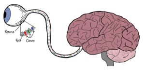
It is in fact the back of the eye which is responsible for the perception and transmission of visual images to our brain. This lining of the eye is known as the retina and is consisted of several, special light-sensing cells. Light stimulation here is then converted into electrical signals that travel all the way to the back of our brain, in the occipital cortex, via the optic nerves and optic tracts.
The retina consists predominantly of two types of cells: rods and cones. Rod cells are stimulated by light over a wide range of intensities and are responsible for perceiving sizes, shapes and movements within the visual field. Cone cells on the other hand, have greater light sensitivity and respond differently to light of different wavelengths, therefore allowing for better colour vision and clarity, so what is otherwise known as visual acuity.
The distribution of rods and cones in the retina isn’t even; overall we have more rod than cone cells, although certain regions are characterised by a greater concentration of cone cells. An example of this is the macula, found posteriorly quite off centred, and in the middle of the macula, the fovea. The fovea is a small, circular region consisting solely of cone cells. This characteristic cellular arrangement allows it to have the highest visual acuity in the whole retinal surface.
For this reason, when we want to focus on an object in our visual field, it is the fovea of the retina that we point at it, allowing our retina to convey a clear and detailed image to our brain of what we are interested in. Everything else which lies outside of the field of vision of the fovea and macula will appear “blurred out” due to the higher density of rod cells on the rest of the retina.
However, this isn’t the only process that takes place when we are trying to focus our vision on a specific object in our visual field. The process is more complex than that and a number of different eye components, including the ones mentioned above, come into play to help us achieve that.
For example, when the object we are trying to focus on is near, our ciliary muscles will loosen allowing for the lens to thicken, while our pupils constrict, and the sclera rotates inward. The opposite process will be true if the object is further away.
Ultimately as we can see (pun not intended) although apparently effortless, the mechanism through which our eyes help us focus on objects in our visual field is much more complex and finely regulated by imperceptible, yet precise, movements than we would expect!


Visit
What's on
Learn and play
Youth Scheme
Quick links
Website by Hut Six Digital